Categories
Emotional Parents

Back to school with an external fixator

Your child must gradually learn to move around with ease doing things he/she used to enjoy. He/she must also learn to be unashamed of his/her orthopedic external fixator. It’s very important for the limb’s healing that your child starts mobilizing soon: he/she must go back to school as soon as safely possible. There he/she can meet school friends, start studying again and get back to a normal routine.

A recovery timeline can be discussed with surgeons and from there you can let school staff know what is happening. They will then be able to prepare for your child coming back to school as well as providing any work that your child has missed while having surgery.

It is important that you and your child talk about his/her external fixator with teachers and friends. This will make school staff and school mates more aware of your child’s special needs regarding extra space, mobility limitations, and rest elevating the affected limb.

Categories
Treatments Parents Safety

Travelling with an orthopedic fixation device

Before deciding to go on a trip with your child, it is important you speak to their health care professional. They may not want your child to travel too far away because of the risk of infection, clots or anything that could happen to the fixation device.

If your child’s doctor says it is okay for your child to travel, ask for a letter from your orthopedic surgeon to pass through security at the airport with the fixator in place and/or medicines related to the surgery.

If flying with an orthopedic fixation device, your child should:

– avoid any salty food and avoid alcohol;

– drink water often;

– use a pillow to help support their limb;

– ask the hostess/steward on board for ice in case the limb starts swelling during the flight;

– show the fixator off – it will make people more aware of his/her needs regarding extra space;

– make sure to have adequate medical supplies such as dressings, pain relief, etc.

– when at the airport, get help with wheelchairs, going and coming from the planes. Be patient and allow yourself plenty of time, as the wheelchair person may be late or the gate may be far away;

– if needed, the carrier could decide to have medical staff on board.

If travelling by car, your child should:

– sit in the front with the seat pushed back, or in the back seat with the legs across the seat;

– use a pillow to make the journey more comfortable.

If travelling by bus or by train, your child should:

– choose the aisle seat – it will be most comfortable as he/she should be able to stretch the limb out.

In all cases, elevate the limb during the journey as much as possible and take regular stops to walk around.

Categories
Parents Safety

Child safety seat for car security

Motor accidents are the most common cause of death for children ages 4 and 14.

Child safety seats have been shown to decrease car death by 71% for kids under 1 and
by 54% for kids aged 1 to 4 years old.

Please follow the tips listed below:

  • It is mandatory to use child safety seats every time we drive, even if it’s a very short journey.
  • Putting children on your lap is never a good idea – this can cause accidents and make driving very unsafe.
  • The back seat is always the safest place for any child safety seat.
  • All car seats must come with the appropriate safety standard labels. Never use one without your country’s accreditation.
  • For a Safe Car Seat Installation, follow instructions from the manufacturer. Do not try to guess!
Categories
Physical Parents Health and Prevention

How to build strong bones in children

It’s easy to take our bones for granted, but they can break, and they take time to heal – even when we are young. We build almost all our bone density and strength when we’re children and teens. Adults build new bone but more slowly, and over time bones get weaker.

As a parent you can make sure your kids get the three key ingredients for healthy bones: calcium, vitamin D and exercise.

Calcium can be found in dairy products, beans, cereals, some nuts and seed,s and leafy green vegetables. Encourage your kids to eat high-calcium foods. Younger kids may need 2–3 servings of calcium-based products each day, while older kids may need 4 servings.

Vitamin D helps the body absorb calcium and can be found in foods such as tuna, cheese, egg yolks, mackerel or salmon, and fortified fruit juices.

Health care providers recommend all kids take a vitamin D supplement if they don’t get enough in their diet. Ask your doctor, nurse practitioner or a dietitian how much vitamin D your child needs and the best way to get it.

Your child’s muscles get stronger the more he/she uses them. The same happens with bones.

Walking, running, jumping and climbing are good for building strong bones, as muscles and gravity put pressure on them. Riding a bike or swimming are great for overall body health, but kids also need to do some kind of weight-bearing exercise for about an hour per day.

Categories
Emotional Treatments Parents

Spotting stress in children

It’s important to remember that kids still experience stress. Things like school pressure, friends and everyday social situations can contribute.

Here are a few ideas of how you can encourage your child to express himself/herself:

– Talk about what you may think is bothering your child, be sympathetic and show you care.

– Listen, be patient and ask your child questions.

– Suggest what you think he/she may be feeling and help him/her process what’s happening.

– Put a label on it to help your child understand his/her own feelings and make him/her realize he/she isn’t alone.

– Suggest ways to tackle the issue together, this will make him/her see that problems can be solved when discussed.

– Limit stressful situations when possible.

– Simply be there, to listen and talk.

– Be patient. You can’t solve every problem, but listening and understanding goes a long way.


What do kids worry about?

Kids and teens can worry about grades, tests, their changing bodies or simply fitting in.
They may feel stressed over social troubles like peer pressure, family problems or whether they’ll be bullied, teased, or left out.

Preteens may worry about world events or issues they hear on the news or at school. Things like terrorism, war, pollution, global warming, endangered animals, and natural disasters can become a source of worry.


How to reduce stress in children?

Here are a few suggestions to help your child:

– Take an interest in what’s happening in and out of school and try to discover if there is a particular issue that’s causing distress.

– Encourage your child to talk about what’s bothering him/her. Sometimes all kids need is a little reassurance.

– Show you care and understand by listening.

– Help them solve the problem or issue themselves and don’t automatically fix it for them. If it is based around world events, take them to a library or encourage them to research to understand more.

– Help your child keep things in perspective.

– Help them see that you’re committed to changing the things they are feeling stressed about.

– Be aware of your own emotional reactions to problems and issues – these can be transferred to kids.

– Be a good role model – your child will mirror your actions.

Categories
Emotional Treatments Parents

How to prepare your kids for the hospital?

Good preparation can help your kids feel less anxious about surgery and get through the recovery period faster. But, like parents everywhere, you’re most likely a little uncertain about the best way to prepare your child.

Explain things in a way that your child understands. Kids of all ages cope much better if they have a good idea of what’s going to happen and why the surgery is needed. Ask your healthcare professionals any questions you have, and do your researches so that you know how to answer questions that your child will ask.

The more you understand, the more your child will too

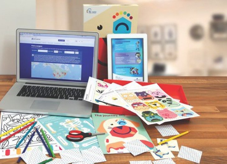
Ask your doctor for appropriate videos or tools that can help explain the procedure.
Find books, appropriate to your child’s level of understanding, about what to expect at the hospital. Reading together and discussing the surgery will make everything seem less threatening.
Remember that as well as the words you use, your tone of voice, expression, gestures, and body language send messages to your child. If you appear fearful, your child is likely to feel fearful regardless of the words you use.

Consider a pre-operative orientation and tour

Hospitals can offer special pre-operative children’s programs, family orientations, and hospital tours. Child-life specialists are an incredible resource for parents and children. They are trained to talk to children about their medical procedures, comfort them if they’re upset or need extra support, and they can even organize group times to get together with children that are going through the same things. An orientation program removes the mystery of the surgery and makes the hospital familiar and friendly and the experience more predictable.

Hospital stays

Many surgeries are now “same-day” procedures with no overnight or prolonged stays needed; many kids are back home the same night.

Anesthesia is much safer today than in the past, but it can still carry some risks. Discuss any concerns you have in advance with the anesthesiologist.

If hospitalization is needed overnight, most hospitals avoid separation anxiety by permitting at least one parent to stay with the child day and night. Check with your hospital about its rules regarding parents staying over and when other close family members can visit.

Explaining the problem and handling fears

Begin by explaining why surgery is needed in simple terms; kids may fear that you aren’t telling them everything, or that their problem is worse than they’ve been led to believe. Build trust and don’t mislead your child – tell as much of the truth as your child can understand.

Kids will most likely be scared that the surgery will hurt. Explain them that a special doctor gives medicine to make them sleep so they won’t feel anything during the operation. Once it is finished, they’ll wake up. Older kids, in particular, may need special assurances that they will wake up.

Explain that you’ll be there when your child wakes up. Tell your child that if anything feels sore right after the operation, a doctor or nurse can give them medicine that will make it feel better.

A teen might be afraid of losing control, missing out on events, being embarrassed or humiliated in public, and sounding childish by expressing fear, anxiety, or pain. A teen also may be afraid of waking up during the operation – or not waking up afterward.

Encourage your teen to read up on their medical condition and share the information with you. Reading and sharing information is an excellent coping mechanism.

A fear of all ages is being seen naked and having their “private parts” touched. If the operation involves the genital or anal area, explain that although it might be embarrassing, doctors and nurses will need to examine these areas, to check if they’re healing after the operation. Be sure to explain that doctors, nurses, and parents are the only exceptions to the rules about privacy.

Children sometimes believe that their medical problem and operation are punishments for “being bad.” They may feel guilty and believe that they’ve brought events on themselves.

Explain that the medical problem is not the result of anything they have done or failed to do, and that the operation is not a punishment, but simply the way to “fix” the problem and that they will feel so much better afterwards.

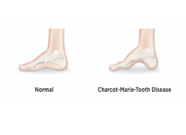
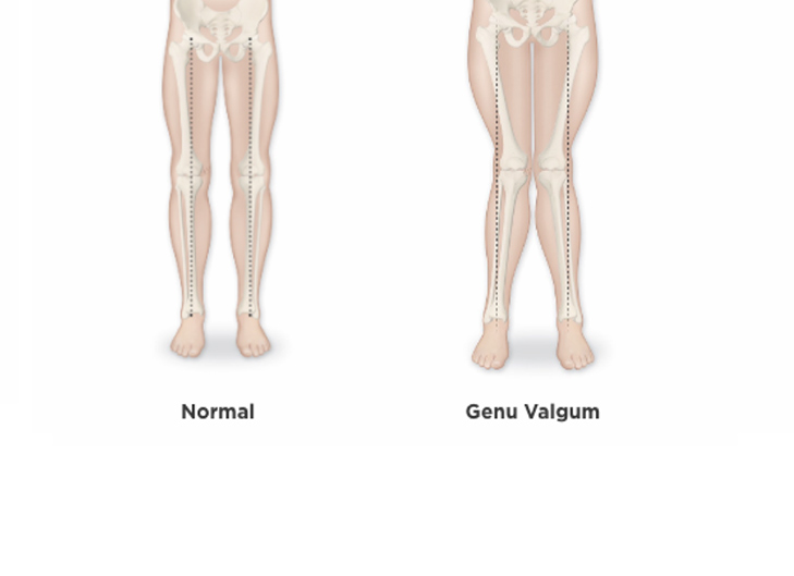
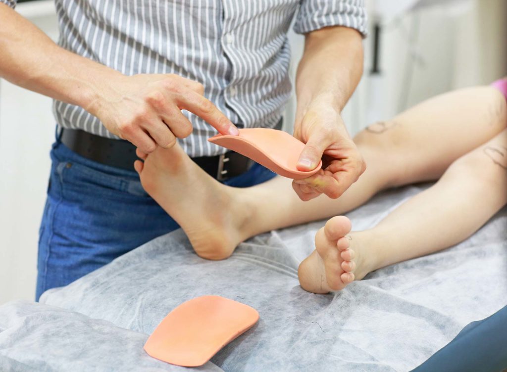
On the day of orthopedic surgery

When you arrive on the day of surgery, your child will have time to wait, so its recommended you bring books or toys from home to pass the time.

After surgery you’ll be allowed in the recovery room to be with your child as he or she awakens.
Distracting your child, whether with a new book or a visit from a relative or friend, can also make recovery more pleasant. Just make sure your child gets plenty of time to rest and recover.

Categories
Physical Treatments Parents

Guide to use of crutches

Getting around on crutches can be tricky at first, but with a few helpful pointers it can be easy and less of a hindrance as you may think.

How to choose the size of crutches?

Ensure the crutches are properly sized so they do not cause problems. Don’t assume the crutches you have at home are the right ones, they may need adjusting.

Proper crutches should be one to two inches below your armpit when you are standing straight. The handles should be at hip height, so that your elbows are slightly bent when holding them.

Check padding and grips of the crutches

Crutches should have ample cushioning on the armpit and grips where the crutches contact the floor.

How to get up from a chair with crutches?

Place both your crutches in the hand on the affected side and grasp the hand rest of a chair with your other hand. Place your weight on your uninjured leg and push up with your arms.

Walking with crutches

Both crutches should move together a short distance in front of you (approximately 18 inches).
Always ensure you take short steps when using crutches. Support yourself with your hands and allow your body to swing forward as if you were going to step on the injured leg; instead of placing weight on the injured leg, rest your weight on the crutch handles.

When going up and down the stairs, go one step at a time, and rest at each step – it’s not a race!

Going up the stairs 1

Stand close to the step, place the crutches at ground level. With your weight on the crutches, pick the uninjured foot up to the step, and then bring the crutches up to the step level. Repeat this process for each step.

Going up the stairs 2

If the stairs have a handrail try holding onto it – with both crutches in the other hand – only if you feel confident to do so.

Going down the stairs with crutches – if you can’t bear weight on your injured leg

If you cannot bear to have any weight on the injured leg, you will need to hold the foot of the injured leg up in front and hop down each step on your good leg. Be sure to support yourself with the crutches or get someone to help you.

Going down the stairs if you can bear some weight on the injured leg

If you can bear weight on the injured leg, place the crutches on the next lower step and step down with the injured leg. Then quickly bring down the good leg. But remember to take it one step at a time.

Don’t let your armpits rest on the crutches, even when resting!

Categories
Emotional Physical Parents Health and Prevention

Motivating teenagers to exercise

The important thing in motivating teens is discovering something that is really fun for them. Not everyone is born to be an athlete, so it’s important to find out if there are some activities that aren’t seen as exercise or competitive and are simply ways of enjoying everyday life.

Never push your child into a sport simply because it’s what you did. Instead, try to talk through what things are interesting to them. It might be that solo pursuits like running or cycling are a better fit than team sports like football.

Competitive sports can add extra stress and pressure, and not everyone thrives in that environment. However, if your child shows a particular skill or aptitude and does like a competitive environment, encourage them. Team sports are great for exercise and building personal skills.

Think outside the box

Try to look beyond the typical forms of exercise or sport. Yoga can be a great way to ensure flexibility and well-being. Going to the gym or swimming might be more interesting to your child. Rock climbing or hiking are great ways to see the great outdoors. Anything requiring movement promotes a healthy lifestyle.

The main point to remember is to listen to what your teenager is interested in and support him/her in whatever exercise he/she feels comfortable with.

Categories
Emotional Physical Parents Health and Prevention

How to make your child active?

From an early age it is important that your children are encouraged to play, exercise and explore. This builds confidence, social skills and physical development.

Regular exercise can prove to be vital in later life, promoting:

    • strong muscles and bones;
    • healthy weight;
    • decreased risk of developing type 2 diabetes;
    • better sleep;
    • a better outlook on life.

It’s proven that active kids are also more likely to be motivated, focused, and successful in school.

Motivate kids to play

The main advice is to keep it fun and light hearted – no child wants to participate in physical exercise if it isn’t fun. Support your child and try to choose things he/she would enjoy rather than just endure. Make it a fun game.

Choosing age-appropriate activities

Try incorporating physical activity into a daily routine. Toddlers and preschoolers should play actively several times a day. Children aged 6 to 17 years should be doing 60 minutes of physical activity daily.

Age-based advice

Younger children need games and exercises that helps them develop motor skills – kicking or throwing a ball, riding a bike with training wheels, or running obstacle courses can all be good.

Some sports may be open to kids as young as 4, although organized team sports are not recommended until children are slightly older. Sometimes kids can’t understand rules and can lack the attention span, skills, and coordination needed to play, but they should still be encouraged to do some form of activity. Remember not to take it too seriously; the fact that they are running around being active is what matters.

School age children can often spend too much time in front of a TV or on electronic devices, so the challenge for parents is to find something they would rather do.

Talking and understanding what they may be interested in is key to identifying the right kind of sport to suggest.

Teenagers have a huge range of choices when it comes to being active: football, running, cycling, baseball, skateboarding, hockey – all of which are great ways of staying active and are normally available to participate in at school or on weekends.

Understanding your child’s attitude to sport

  • Non athlete

General lack of athletic ability, interest in physical activity.

  • Casual athlete

Generally interested in being active but is at risk of getting discouraged in a competitive athletic environment.

  • Athlete

High athletic ability, committed to a sport or activity and likely to excel with the right support.

Regardless of which category your child falls into, be positive and support any form of exercise that your child shows an interest in – it all helps promote a healthy lifestyle.

Categories
Emotional Physical Parents Health and Prevention

How to keep your kid active?

Kids love to play and it’s an essential part of their growth, so it should be encouraged. Not only should their body be active, but their brains also need stimulation.

Don’t let young children spend extended periods of time in high chairs, strollers, and car seats. Screen time on televisions or electronic devices should also be limited.

Playing with toys is a much better way of keeping minds and bodies active.


Here are some ideas to keep toddlers active at home:
12-24 months

-Listen to music and encourage dancing together

-Jumping – while holding your toddler’s hand

-Climbing stairs with supervision

-Playing with building blocks and other physical toys

24-36 months

-Play at the playground

-Gentle ball games

-Toy cars

-Building blocks


Investigate how active your kids are away from home supervision, for example:

– Do they go outside most days?

– Is there a schedule of activities they adhere to?

– Do they watch videos or TV, and if so, how much and how often?

Think about joining a playgroup. Playgroups offer a change of pace.


Safe environments and playgrounds

Play areas must be safe, so always check that a playground has age appropriate equipment that is clean and well-maintained. Some children do not have any fear and will attempt things that could be dangerous. To reduce the risk of injury and make playing more fun for everyone, stick to play areas that are designed for your child’s age.

Categories
Emotional Physical Parents Health and Prevention

How to encourage children that don’t like sports to exercise?

Team sports can be great to keep kids fit and help them with self-esteem, learning about teamwork and making friends, but sometimes children can be a little reluctant to participate.

Some kids find it difficult to take part in team sports for a variety of reasons. Shyness, fear of not being good enough, or worrying about looking foolish in front of friends are feelings that can all impact your child’s interest in sports.

Kids are still developing basic skills

It isn’t until around the ages of 6 or 7 that kids start to develop the physical skills needed to effectively play sports. One thing you can do to help them is practice: the more your child plays sports with you and friends at home, the more confident he/she will be in a competitive setting. It also means you will spend quality time together!

Your child may enjoy playing sports for the fun of it, but doesn’t want to compete. Listen to your child. He/she may be more likely to enjoy the sport if it’s just played for fun.

Make the right choices when it comes to skill level

Try to assess your child’s skill level when selecting teams or sports. It’s not a good idea to push your child into a sport if he/she isn’t ready. Many sports have beginner level classes, and can be the best place to start when your child is still learning the basics.

Fight or flight

Some kids aren’t natural athletes. It’s important to provide positive encouragement and support from the start so they understand that it’s okay to make mistakes. Learning a sport takes time, and everyone makes mistakes.

Choosing the right activity

Have an open mind when selecting the right sport for your child. Some kids struggle with coordination, so sports like cycling or swimming might be a better option than football or baseball.

Other factors

Many factors impact a child’s ability to participate in sports. They may be overweight, have asthma, or some other health issues that make it difficult for them to keep up. Try to select a sport that is appropriate for your child’s attitude, skill level, and interests. If your child doesn’t enjoy a sport, they won’t want to do it.

When team sports aren’t right

If a team sport isn’t a good fit for your child, here are some individual activities to try:

  • swimming
  • horseback riding
  • dance classes
  • inline skating
  • cycling
  • cheerleading
  • skateboarding
  • hiking
  • golf
  • tennis
  • fencing
  • gymnastics
  • martial arts
  • yoga and other fitness classes
  • running

Be patient

Sometimes finding the right sporting activity can take time and patience. It’s important to find an activity that feels like a hobby and not a chore. The reward of finding the right activity for your child and promoting a healthy lifestyle will be well worth it.

Categories
Treatments Parents Health and Prevention

Bone deformity correction: when is it time for Health Care Provider consulting or check-ups?

Human skeletal development starts during pregnancy and continues until bones have reached their full shape and structure in late puberty.

Some bone deformities are evident at birth, such as clubfoot, while others generally occur between 10 and 18 years old, such as scoliosis where the spine has a side-to-side curve, with an S or a C shape.

If your child shows signs of a bone deformity, you should take them to your Health Care Provider as soon as possible for an assessment. The earlier a bone deformity is diagnosed and treated, the more successful the treatment will be.

Your HCP and/or orthopedic surgeon will carefully evaluate what is appropriate for your child, considering the following aspects: your child’s age; her/his medical and family history; a physical examination; imaging, and blood and/or nerve tests.

It is important that you feel comfortable asking your HCP any questions about your child’s diagnosis and treatment that you may have. The better informed you are, the more comfortable you and your child will be.

Categories
Physical Treatments Parents Conditions Bone Fractures

What is a growth plate and a physeal fracture?

Almost every bone in your body has a physis. The physis (or growth plate) is made of cartilage and is near the ends of the long bones. Physis helps bone grow in both length and width. When a child stops growing, the physis hardens into solid bone.

Physeal fractures are very common; the most common types of fractures are found in the fingers, wrists and lower leg, mostly from overuse and in playing sports. The physis can fracture and although physis can heal very quickly, it’s important to bring your child to an orthopedic specialist as soon as possible after an injury occurring.

Physeal fractures classification

The Salter-Harris classification describes the different patterns of physeal fracture:

Type I fractures
This could just be a crack in the physis that is still perfectly aligned, or the physis may be disrupted and the bone separated.

Type II fractures
A fracture that is partly through the physis and then out the shaft of the bone away from the joint.

Type III fractures
A partial fracture through the physis and then exits the end of the bone into the joint. The joint surface may be disrupted.

Type IV fractures
A fracture that goes through the shaft of the bone, also through the physis, then exits out the end of the bone at the joint.

Type V fractures
Part of the physis is crushed but not displaced; these can be very difficult to see on X-rays.

Physeal fracture symptoms

Swelling, pain, bruising, and a crooked limb with the inability to move are the most obvious signs that a physeal fracture has occurred.

Physeal fracture examination

A doctor will need to test the joints and force some gentle movement to diagnose the problem. They will then conduct X-rays, MRI or CT, which can help to correctly identify a physeal fracture.

Physeal fracture treatment options

Treatment will depend on the specific injury. Treatment options include a cast or surgery using metal pins or screws.

Physeal stress fractures can often heal with rest, although a splint or cast may be used to help with the pain. Following healing, it’s important to reduce stress on the affected area and avoid any activity that could cause further injury or stop the healing process. Your surgeon will give you guidelines on this.

Physeal fracture surgical treatment

Surgery may be needed if:

  • the skin is cut near the broken bone;
  • the fracture will not stay lined up even with a cast;
  • bones have started to heal incorrectly.

Implants can be used in surgery to keep the bones aligned properly. This can include plates, pins or screws to help keep the bones aligned while healing.

Outcome

Growth plate fractures, especially in the femur (upper part of the knee), must be monitored for 9 to 12 months after injury to ensure the bone grows properly. X-rays are needed to monitor progress.

If a hard-bony area forms across the physis, it may stop growing properly or grow in a crooked form. This is very rare, but if it does happen, there are different techniques for removing the bony bridge, including inserting some fat into the area, or cutting the bone to adjust its growth pattern. Your doctor may suggest surgery to prevent this from happening.

Categories
Treatments Parents Conditions

What is Pes Planus?

Pes Panus deformity is more commonly known as flatfoot. Both children and adults may be flat-footed, and it’s estimated that the prevalence of Pes Planus in the general population is about 20-30%.

What causes Pes Planus?

The foot must act as a reliable landing point when putting the foot down, usually heel first, and as a rigid taking off point when walking or running. In pathological flatfoot, the arch of one foot or both feet have either not developed normally, or an injury or condition has produced it, such as rheumatoid arthritis, stroke or diabetes. It may develop as a result of degenerative changes in joints and/or ligaments; this will tend to be in older patients and people who are very heavy. It may also occur as a result of trauma to the bones and/or ligaments. An increasingly common cause seems to be neuropathic problems secondary to diabetes, as an early manifestation of Charcot foot.

Pes Planus symptoms

The arch of the foot is lowered or flattened out, depending on the severity. Slightly lowered arches are usually symptom free. Issues such as discomfort and chronic pain can arise if there is a minor injury, sudden weight gain, poorly-fitted footwear, and excessive standing, walking, jumping or running.

Pes Planus diagnosis

Pes Planus diagnosis is usually by clinical examination. In young people, the flat foot that is apparent when standing disappears when they stand on tip toe, and the normal arch appears. This is flexible Pes Planus, is usually symptomless and requires no treatment. X-rays may be helpful when there is a history of injury but may not be necessary.

Pes Planus treatment

Management in less severe cases of Pes Planus generally consists of wearing spacious, comfortable footwear with good arch support, possibly supplemented by padding or orthotics in order to balance and cushion the foot. The object of Pes Planus treatment is to restore anatomy and function as much as possible. Orthopedic surgery, sometimes with osteotomy, may be necessary. Bone grafting to replace lost bone may be also necessary. After surgery, your child may need to wear orthotics. Since each case is different and there are many types of treatments, discuss the best treatment for your child with your doctor and orthopedic surgeon.

Pes Planus deformity complications

If not treated, some cases of Pes Planus can result in severe deformity and early onset of arthritis of the foot and ankle. This may cause severe pain and significantly reduce the ability to walk, even when wearing orthotics.

Categories
Treatments Parents Conditions

What is Pes Cavus?

Pes Cavus is another name for a high-arched foot. Prevalence in the population is estimated to be 8-15%.

What causes Pes Cavus deformity?

Severe Pes Cavus can be caused by progressive neurological disorders (e.g., spinal trauma, muscular dystrophy, hereditary neuropathy), static neurological disorders (e.g., stroke, cerebral palsy) and other causes, such as foot trauma.

Pes Cavus disease symptoms

The shape of the foot may range from a slight high arch to a severe deformity that causes a patient to walk on the outside of the foot.

Pes Cavus diagnosis

An X-ray will confirm a diagnosis of Pes Cavus and its severity.

How to treat Pes Cavus?

Slight high arches are usually corrected or managed with specially designed footwear (orthotics) and severe deformity may require orthopedic surgery to achieve realignment. Osteotomy (the surgical cutting of a bone or removal of a piece of bone) and an external fixation device can correct a severe bone deformity, thus reducing pain, improving function, and decreasing the incidence of deformity-related injuries, such as ankle sprains and broken bones. However, realignment through an external fixator must be supplemented with soft-tissue-balancing procedures, such as tendon transfer and orthotic management, in order to maintain the correction.

Pes Cavus complications

Left untreated, Pes Cavus causes foot pain, and potentially knee, hip and back pain that limits mobility. People with Pes Cavus deformity may also be more prone to broken bones in the lower legs.

Categories
Treatments Conditions Parents

What is Charcot-Marie-Tooth disease?

Charcot-Marie-Tooth disease (CMT) is a group of inherited neurological disorders involving the peripheral nerves. Peripheral nerves are outside the brain and spinal cord, and govern the motor and sensory capabilities in the limbs (such as walking). CMT’s estimated prevalence in the general population is 1 out of 2,500 (0.04%).

CMT is caused by congenital mutations in genes involved with the structure and function of the nerves governing feet, legs, hands and arms. There is no single faulty gene and there are many varieties of CMT caused by different genetic faults.

Charcot-Marie-Tooth disease symptoms

CNT usually develops between 5 and 15 years old, but it may develop later. Foot deformities may occur, and the lower legs may have an “inverted champagne bottle” appearance as muscle bulk is lost. As the disease gradually progresses, weakness and loss of fine motor skills may occur, with pain ranging from mild to severe.

Charcot-Marie-Tooth disease diagnosis

A neurologist can diagnose CMT after nerve conduction studies, electromyography to measure the electrical activity of muscles, and sometimes by nerve biopsy.

Charcot-Marie-Tooth disease treatment options

Although Charcot-Marie-Tooth disease cannot be cured, it can be managed with physical therapy, braces, pain medication and surgery. Osteotomy and treatment with an external fixation device can help to reverse foot and joint deformities, aiding support and strength.

Charcot-Marie-Tooth disease complications

Left untreated, CMT leads to pain and difficulty walking, which decrease everyday activity levels and adversely affect quality of life. Foot wounds, foot fractures and more-severe deformities of the foot and ankle may also occur.

Categories
Treatments Parents Conditions

What is angular deformity of long bones in growing children?

Angular malalignments of long bones are a common concern in the early years of life. In most cases, symmetrical deformities and absence of symptoms present a benign condition with excellent long-term outcome.

On the contrary, deformities that are asymmetrical and associated with symptoms may indicate a serious underlying cause leading to aesthetic and functional deficits that will require surgical orthopedic treatment.

At what age can angular deformity of bones be considered a pathology?

Genu varum (bowlegs) are normal in newborn and infants up to 12 months. It improves with growth: when child starts standing and walking the lower limbs gradually straighten (with normal growth) and the genu valgum spontaneously correct at the age of 7. If they persist after the age of 10, they may require surgical orthopedic treatment.

Angular deformity causes

Some experts think that a high amount of stress and strain imposed too early on a joint, during growth and/or adolescence (for example, through intensive sport practice) may contribute to this type of growth deformity, but so far not enough scientific data is available. However, it’s known that the most frequent causes of angular deformity of long bones in children are growth disorder, previous trauma or injuries, cancer and/or infections, or a pathologic condition – often caused by Blount’s Disease (tibia vara) and renal rickets, or vitamin D deficiency.

Angular deformity diagnosis

Familiarity with the natural history of angular deformities and with normal growth patterns is necessary to diagnose and evaluate malalignment. Diagnosis should include family history, any description of onset and information about the progression of the bone deformity, plus a selective use of X-rays, CT and MR imaging. The child will be observed walking, with attention to her/his knees during the stance phase, to determine if lateral thrust (genu varum) or medial thrust (genu valgum) occurs. The doctor should know the diet and amount of vitamins taken by the child, inquire about milk allergy, or intoxication of metal – specifically to lead and fluoride.

Angular deformity treatment options

Treatment options depend mainly on the degree of bone deformity and the age of the child. Treatment is rarely required if the child is under 18 months. In case of moderate, milder deformity (stage I and II), this can be treated with a night time orthosis, and/or a day time brace, which can be effective up to the age of three years.
All children with significant angular deformity (stage III-V) should be considered for orthopedic surgery by osteotomy with an internal and/or external fixation device. If performed at an early stage, the surgeon will usually obtain complete and permanent correction of the child’s limb deformity, whereas there is a greater chance for recurrence if osteotomy is done in later years.

 Possible post-surgical complications

Bone lengthening and deformity correction can come with a few possible complications. The most common are the following:

  • infection – for instance, with an external fixation device it may occur at the pin and wire sites (which must be constantly cleaned for prevention), or in the bone (which is extremely rare);
  • delayed or too fast bone healing;
  • implant related complications;
  • nerve or blood vessel injuries;
  • muscle stiffness and joint contracture;
  • bent or broken bones;
  • blood clots in the leg;
  • allergies to some medications, or anesthesia.

Ask your doctor or orthopedic surgeon to explain all the possible post-surgical complications and how they can be prevented. Do not hesitate to contact her/him whenever you have any doubts or questions.

What can happen if a bone deformity is not corrected in time?

If left untreated, or not treated in time, bone deformity can deteriorate with age and lead to major complications. While growing, your child will gain weight, which adds extra stress to his/her joints. The bone deformity can become permanent and in time your child can develop osteoporosis, with further reduction of the mobility and more fragile bones. There can also be increased severe functional and aesthetic problems, nerve damage and pain.

Ask your doctor or surgeon to explain all the possible complications if your child’s bone deformity is left untreated. Contact her/him for any doubt or question you may have.


Categories
Treatments Parents Conditions

What is Arthrogryposis Multiplex Congenita?

`Arthrogryposis is a non-progressive – it does not worsen over time – disorder that is present at birth, involving many muscle and tendon shortenings that limit joint movement. It occurs in approximately 1 out of 3,000 births (0.03%), and can affect all four limbs, or upper or lower limbs only.

Arthrogryposis causes

It is present at birth and the exact cause is unknown.

Arthrogryposis symptoms

Muscles and tendons are shortened, so the limbs may be stuck in one position. The child’s limbs are often thin, with weakened muscles and abnormal joints resulting in, among others, knee contractures and foot conditions.

Arthrogryposis diagnosis

A doctor will perform an examination, taking into account the symptoms and their appearance. If more information is needed, imaging tests – such as X-ray, CT scan or MRI – may be performed, along with more specialized tests, like muscle or skin biopsy, blood tests and nerve tests.

Arthrogryposis treatment options

Arthrogryposis can be treated with joint manipulation, orthotics and casting, particularly in the first few months of life. For correction of severe and ongoing deformities, surgery may be required. Surgery can be as simple as releasing the Achilles tendon or the use of an external fixation device, which may be required for realignment and to lengthen limbs that have contractures.

Arthrogryposis multiplex congenita complications

Proper treatment helps improve the range of motion and your child’s ability to use their limbs, although there is no way to completely fix arthrogryposis. Depending on the severity of the condition, people with arthrogryposis may have few physical limitations after treatment, while others who are more severely affected may live with discomfort and lack of mobility, and require physical assistance for daily activities.

Categories
Treatments Parents Conditions

What is Genu Valgum?

Genu valgum, also known as knock knees, may be a passing trait in children. It can be seen in children from 2 to 5 years old and is often naturally corrected as children grow. When knock knees are severe, there can be strain on the knee, which results in pain.

What causes genu valgum?

Genu valgum can be passed down through genes, or it can happen because of injury, infection or a problem with metabolism that has affected the bones. It can be caused by poor nutrition – by obesity and, less commonly, by severe calcium and vitamin D deficiencies.

Genu valgum symptoms

When standing, your child’s knees will touch or be closer together than the ankles, pushing the ankles further apart. Knock knees can cause pain, discomfort, a limp or difficulty walking.

Genu valgum diagnosis

An orthopedic specialist will look at the legs to see if the angle is not in a normal range. An X-ray of the knee may be needed to confirm the severity of knock knees.

Genu valgum treatment methods

This deformity can sometimes be managed with noninvasive methods, such as limits on activity, nonsteroidal anti-inflammatory drugs like ibuprofen, braces, exercise programs and physical therapy. When these methods don’t work, surgery may be needed. Osteotomy (the surgical cutting or removal of a piece of bone) and correction with external fixation is a useful way to support and straighten the limb in the management of genu valgum. Guided growth may be a good treatment in some cases.

Genu valgum complications

If untreated, the symptoms can worsen, causing pain, problems with movement and even arthritis.

Categories
Treatments Parents Conditions

What is Genu Varum?

Genu varum, or bowlegs, may present at any time from infancy through adulthood. It can affect one or both legs. Medical and genetic history may help doctors understand how the condition will progress and for how long. As genu varum becomes more severe, the patient may start to waddle and have discomfort while walking. In children up to 2 years old, painless bowing on both sides may occur and often fixes itself over time.

What causes genu varum?

Genu varum is normal in the very young and may happen in older children due to rickets and Blount’s disease, or because of bone problems, infections or tumors.

Genu varum symptoms

One or both legs may show bowing, often just below the knee, even when the ankles are together. It may progress rapidly and unevenly.

Genu varum diagnosis

An orthopedic specialist will look at the legs to determine how far they bend inward and will confirm the diagnosis with an X-ray of the knee.

Genu varum correction treatment options

Bow-leggedness that causes problems and does not fix itself will be seen in X-rays and may need treatments, including surgery. Physical therapy exercises, special shoes and limits on standing and activity may be needed before or after surgery. Guided growth may be the best genu varum treatment to help straighten the limb. In certain cases, genu varum treatment with osteotomy (the surgical cutting of a bone or removal of a piece of bone) and an external fixation device is necessary.

Genu varum complications

If severe bow-leggedness is not treated, the symptoms can worsen, causing different leg lengths, pain and difficulties with mobility.

The condition can come back after surgery, especially in younger children, who are still growing. An orthopedic specialist can help you determine the right time to perform surgery.

Categories
Treatments Parents Conditions

What is Rickets?

Rickets is a condition that softens, distorts and weakens bones in children. As late as the 1940s rickets was a very common childhood ailment.

Rickets causes

A lack of vitamin D or calcium is the most common cause of rickets in children. Among the genetic rickets causes – those that cannot be fixed simply by changing diet and nutrition – the most common is X-linked hypophosphatemic rickets, which happens in 1 out of 20,000 newborns (0.01%). Other genetic causes are very rare.

Rickets symptoms

Children with rickets may suffer from bone pain, bones that break easily, stunted growth, muscle cramps and bone deformities such as bow-leggedness (genu varum), and spinal deformities.

Rickets diagnosis

Blood tests can diagnose rickets disease and an X-ray may be needed to understand the extent of bone deformities. Bone density scans may be needed to see how severe the disease is.

Rickets treatment options

In case of deficiencies, the first line of rickets treatment is to add vitamin D and calcium to the diet, to let the body fix any bone problems itself. However, rickets disease caused by a genetic condition may need additional medicines and surgery. In less-severe cases, a brace can be used on the spine or limbs to support the bones as they grow.

In more severe cases of limb deformity, gradual osteotomy (the surgical cutting or removal of a piece of bone to change how bones line up) and the use of an external fixation device can be used to both support and reshape the limbs as they grow and get stronger. Guided growth with small plates can be used to correct deformities.

Rickets disease complications

If untreated, rickets can lead to very stunted growth, dental problems and seizures. Bone problems need to be treated to prevent pain and issues with mobility.

Categories
Treatments Parents Conditions

What is Clubfoot?

Clubfoot is diagnosed when a baby’s foot is twisted down and in. Usually present at birth, infant clubfoot occurs in about 1.3 out of 1,000 births (0.13%) and is usually an isolated problem for an otherwise-healthy baby. About half of children with clubfoot have the condition in both feet.

Infant clubfoot causes

The cause is at least partly genetic, because it is known to run in families. Otherwise it is not known, but it might happen when muscles are not even in the lower leg.

Infant clubfoot symptoms

In clubfoot, the top of the foot usually twists down and in. The arch is high, and the heel also turns in. In more-severe cases, the foot might actually look upside-down. Even though clubfoot may shorten the foot and affect normal growth of the calf muscles, clubfoot itself doesn’t actually cause discomfort or pain.

Infant clubfoot diagnosis

A doctor can usually diagnose infant clubfoot based on how the foot looks after birth or sometimes also during routine ultrasound scans during pregnancy. In some cases, an X-ray may be needed to determine the severity of the condition.

What are clubfoot treatment options?

Clubfoot hinders normal walking so treatment should begin soon after birth. The most common clubfoot treatment method involves stretching and casting (to hold the new position of the foot), performed repeatedly over several months, followed by minor surgery to lengthen the Achilles tendon under local anesthetic (Ponseti method). Stretching and use of special shoes and braces may be needed for up to three years after the surgery. Most children treated this way will have pain-free, normal-looking feet that function well.

However, severe clubfoot that has not gotten better with stretching and casting may need surgery and possibly the use of an external fixator, which can be used to help reshape muscles and other soft tissues over the course of several months to one year. In most cases, babies who are treated early are able to wear ordinary shoes and participate in normal activities when they are older.

Clubfoot complications

Even after clubfoot treatment, once your child starts to stand and walk, mobility may be slightly limited. In addition, shoe sizes may differ between the clubfoot and the unaffected foot. If left untreated, your child is likely to have a lack of normal muscle growth, arthritis, inability to walk normally and, perhaps, self-esteem issues related to how the foot looks.

Categories
Treatments Parents Conditions

What is Blount’s Disease?

Blount’s disease, also known as tibia vara, is a condition in which growth does not occur normally in the growth plates of the lower leg, resulting in deformities that may differ in direction and severity. Severe bowing of the legs can result, causing pain and mobility issues. It is different from physiological bow legs, which tend to straighten as the child grows. Your doctor will be able to tell you which one your child is experiencing.

Blount’s disease causes

The cause of tibia vara is not certain, but it seems to be associated with early walking and above-average weight, and may be caused by the effects of weight on the growth plates. There is a genetic element and some patient groups seem more likely to develop Blount’s Disease.

Blount’s disease symptoms

One or both legs may show bowing, which typically occurs just below the knee and may progress rapidly and unevenly.

Blount’s disease diagnosis

An orthopedic specialist will examine the legs to determine the inward-bending angle and will confirm the diagnosis by performing an X-ray of the knee.

Can Blount’s disease be cured?

In younger children and less severe cases, a corrective leg brace or orthotic may be used to manage the limb deformity. Another option is a minimally invasive orthopedic surgical procedure to insert a guided growth plate system. This inhibits the bone growth on one side of the deformity and allows the opposite side to catch up, straightening the leg.

In more severe deformities, an osteotomy may be required. After osteotomy, either an external or internal fixation device will be used to hold the new bone in place.

What are Blount’s disease or tibia vara complications?

If Blount’s disease is not treated, the symptoms can get worse, causing different leg lengths, pain and trouble walking. The condition can come back after surgery, especially in younger children, who are still growing. An orthopedic specialist can help you determine the right time to perform any surgery.

Categories
Treatments Parents

How to correct bone deformity?

When a limb needs to be lengthened, or a bone needs to be corrected, there are several orthopedic non-surgical and surgical treatments available, depending on the severity of the issue and the patient’s medical history. Your doctor or orthopedic surgeon will carefully evaluate what is appropriate for the case considering your child’s age, her/his medical history, the physical examination, imaging, and blood and/or nerve tests.

What is the most common bone deformity surgical treatment?

The most common surgical method to treat bone deformities consists of carefully cutting the bone on purpose and then lengthening or correcting the limb. This corrective procedure, called osteotomy, may be performed with either internal or external fixation devices (more rarely together) to stabilize the cut bone.

Orthopedic internal fixation allows to correct the bone deformity all at once. The surgeon will operate your child under general anesthesia, and will apply metal rods, screws or plates – that will remain in place under the skin after orthopedic surgery – to adjust the bones.
Orthopedic external fixation, with metal rods or pins located outside the limb, is recommended in case of a quite complex bone deformity, which cannot be repaired using open reduction with an internal fixation device, and which is safer to correct gradually, avoiding injury to soft tissues such as nerves or blood vessels.

How can a bone deformity be corrected with an orthopedic external fixator?

An external fixation device allows the new bone to strengthen and harden while growing in the new shape/length. To achieve the bone deformity correction, the external fixator needs gradual adjustments to help the new bone tissue to grow into place over time. When the orthopedic surgeon is happy with the bone’s length, shape and alignment, adjustments will no longer be needed. However, your child will still wear the external fixator, which will support the limb while the new bone tissue hardens and the limb becomes stronger (Bone healing process). These two phases – the “correction” and “consolidation” steps together – usually last from 4 to 12 months.

At the end of the bone fracture healing process, the external fixator will be removed. Your child may need to wear a cast for a short while after the fixator frame has been removed.

What is a hexapod-based system?

A hexapod-based system is a new and advanced method for bone deformity correction. It combines two parts: hardware and software. The hardware is made of metal rings that are anchored to the bone by screws and wires. The software incorporates imaging to determine the precise measuring and positioning of the fixator and allows for its simultaneous adjustment at multiple angles. This innovative system lets children with bone injuries/deformities receive customized care that can optimize clinical, aesthetic and functional outcomes.

How can a bone deformity be corrected with guided growth plates?

In children, bone deformity can gradually be corrected by stapling one side of the growth plate. By doing this, one side of the growth plate is harnessed, while the other side goes on growing. As soon as the bone is straight, the staple can be removed to allow symmetric development of the whole growth plate. This orthopedic surgical technique has the advantage to be minimally invasive and it is often performed on an outpatient basis.

Categories
Parents Conditions

What is a bone deformity?

A bone deformity is an atypical, structural deviation or distortion of the bone’s shape from its normal alignment, length and/or size. A bone deformity can be congenital or acquired.

What causes bone deformities?

Children’s bones grow and reshape themselves continually and extensively. Growth proceeds from a very vulnerable part of the bone named the growth plate (What’s the difference between Kid bones and Adult bones?). While reshaping and remodeling, old bone tissue is replaced by the new one, and many bone disorders and/or deformities come from these changes. This type of deformity is called developmental – caused by the changes that occur in a growing child’s musculoskeletal system. These deformities may get better or worse as children grow.

Bone deformities can also be:

  • Congenital – they are inherited, sometimes related to specific pathologies.
  • Post-traumatic – they occur after accidents and injuries, when the bone heals in a deformed position.
  • For unknown reason – they just happen. In this case they are called idiopathic.

How many types of bone deformity are there?

There are four main types of bone deformity:

  • a bend in the bone – called “angulation”
  • a twist in the bone – called “rotation” or “torsion”
  • a shift in the position of the bone caused by a fracture or by osteotomy – called “translation” or “displacement”
  • a difference in the length of a bone compared with the contralateral – called “limb length discrepancy”.

All these different types of bone deformity can exist on their own, but it is common to find a combination of them.

Categories
Treatments Parents Bone Fractures

How to provide first aid treatment for a bone fracture?

In case of bone fracture, you should:

  • Call immediately for medical care.
  • Keep your child still and calm.
  • Do not move your child in case of head, neck, back, pelvis or hip injury.
  • Support the injured limb with a pillow or sling.
  • Check your child’s airway or breathing.
  • If skin is broken, it should be treated to prevent infections. If possible, rinse the wound gently to remove visible dirt and cover with sterile dressing.
  • If needed, immobilize the broken bone with a splint: these may include a rolled-up newspaper, cardboards or strips of wood. Immobilize the area both above and below the injured bone.
  • Apply ice packs wrapped in cloth to reduce pain and swelling.
  • Prevent your child’s shock. Lay her/him down, if possible, with elevated limb about 12 inches-30 centimeters higher than the heart.
  • Do not move your child in case of head, neck, back, pelvis or hip injury.
  • Treat bleeding by placing a dry clean cloth over the wound to dress it.
  • Pain relief may be needed and medication may help to reduce pain. Follow dosage instructions given by your doctor.

In case of bone fracture, you shouldn’t:

  • move your child unless the broken bone is stable;
  • move a child with a suspected injured spine, head, neck;
  • move a child with injured hips, pelvis or upper leg, unless absolutely necessary;
  • attempt to straighten a bone;
  • apply heat in any form, since it increases swelling and pain;
  • allow your child to eat anything, in case surgery is needed.

Which are the exams for a correct diagnosis?

X-rays are generally used to diagnose the type of bone fracture and whether or not the bones are in line (if there is a displacement or not). Although X-rays reveal most fractures, including subtle fractures in skeletally immature children, in some cases (e.g. occult physeall fracture) the fracture detection may be improved by using magnetic resonance imaging (MRI) or computed tomography (CT or CAT scan).

Does physiotherapy help my child’s bone fracture healing?

Physiotherapy exercises are very important for your child’s limb healing. Physiotherapy makes sure that his/her bones are surrounded and supported by healthy, strong muscles, and his/her joints continue functioning well to prevent permanent joint stiffness. Exercises must be constantly practiced under the supervision of a qualified physiotherapist.

Useful resources to help bone health

The two most important lifelong bone health habits are proper nutrition – a varied and balanced diet with the right amount of essential elements plus calcium and vitamin D – and physical activity. One of the best ways to encourage healthy habits in your children is to be a good role model yourself. Children watch us, and our habits have a strong influence on theirs.

Categories
Treatments Parents Bone Fractures

Types of bone fractures in children

There are several types of bone fractures that may occur in children, including:

  • Greenstick or Buckle – an incomplete fracture in which the bone bends or buckles without completely breaking; frequent in small children.
  • Open (or Compound) – the fractured bone breaks the skin.
  • Closed (or Simple) – the fractured bone doesn’t break the skin or outer tissues.
  • Comminuted – a fracture of three or more relatively small fragments.
  • Displaced – the bone cracks completely in many pieces that move out of alignment.
  • Transverse – a fracture that goes across the bone’s axis.
  • Spiral – a fracture that runs around the axis of a bone.
  • Oblique – a fracture that goes at an angle to the axis.

What are the most frequent bone fracture treatments?

The most frequent bone fracture treatment options are the following:

  • Splint or Plaster Cast.
  • Open reduction with internal fixation (ORIF).
  • Open reduction with external fixation (OREF).
  • Closed reduction with external fixation (CREF).

The needed treatment will depend on the type of bone fracture.

What are the main differences among these bone fracture treatments?

  • Splint and Casting are non-surgical options. Splints are mostly used in cases of incomplete fractures and casts are used for closed, standard fractures. Always follow your doctor’s instructions completely.
  • Open reduction with internal (ORIF) or external fixation (OREF), and more rarely together, are options that require orthopedic surgery. ORIF is a recommended surgical procedure in case of complicated bone fractures not able to be realigned (reduced) by casting, or in case the long-term use of a cast is not desirable or indicated. The orthopedic surgeon will operate while your child is under general anesthesia, and will apply metal rods, screws and/or plates to repair the bones that will remain in place under the skin after surgery. OREF is a surgical procedure that involves the use of an external fixation device to support the bone and hold it firmly in the correct position while it’s healing. The orthopedic fixator is connected to the bones with bone screws, commonly called pins, and will be removed when healing is achieved. OREF is recommended in case of quite complex fractures that cannot be repaired using open reduction with an internal fixation device.

Who decides the best treatment options?

The treatment your child needs will depend on the type of fracture, age, overall health and medical history of your child. Each child is different and your orthopedic surgeon – a doctor specialized in conditions related to bones health, ligaments, tendons and muscles – will discuss with you the necessary and best treatment for your child’s bone fracture. Your orthopedic surgeon will also share with you your child’s recovery plan and will be with you along the entire treatment to ensure the treatment is proceeding as intended.

What to expect after surgery with an external fixator?

In case your child’s limb has been realigned with the use of an external fixator, your orthopedic surgeon will give you an estimate of how long your child’s recovery will take, as it depends on many factors: type of bone fracture, child’s age, overall child’s health and other co-factors. During the healing process, it’s important that your child stays active and starts mobilizing as soon and safely as possible: playing, going back to school, keeping up his/her daily routines. When back home, your child should meet other children and friends, enjoying their company to overcome the initial stress and limitations, and always following the instructions provided by the surgeon and hospital staff.

In case of treatment with orthopedic external fixator, why is pin-site care so important?

The pin-site is the area of skin crossed by pins or wires of the external fixator: it has to be checked carefully every day and kept constantly clean to prevent infections of your child’s limb. It’s very important that you follow exactly the instructions provided by your orthopedic surgeon and hospital staff.

How long does an external fixator stay on?

The hardware will be removed from your child’s limb when the orthopedic surgeon is completely satisfied with the alignment and consolidation of the bone. Your child might need to wear a cast for a short while after the fixator frame has been removed.

Categories
Parents Bone Fractures

Bone fracture healing: how does it work?

Let us start from the basics: what are bones made of? The main component of a bone is bone matrix, which is a mixture of a fibrous protein called collagen and an inorganic compound mostly made of calcium and phosphate. All bones have an outer surface called periosteum, a very thin membrane with nerves (that feel pain) and blood in it. The compact bone is the smooth and very hard part of the bone; it’s what we see when looking at the skeleton. The spongy bone is lighter than the compact bone, but still very strong. The inside of a bone is hollow,and filled with a jelly called bone marrow.

What’s the difference between kid bones and adult bones?

  • Your baby has tiny hands, little feet, small limbs and an overall tiny body. As he/she grows up, everything becomes gradually bigger, including his/her bones. An infant has about 300 bones at birth; while growing, they fuse together to form the 206 bones that an adult has. Some of your baby’s bones are totally made of a special, soft and flexible material called cartilage; some others are partly made of cartilage. Cartilage is replaced by hard bone matrix as the bones reach their full size. By the time he/she is about 25 years old, this process will be complete and the bones are as big and long as they will ever be. All together they form a skeleton that is light but also strong and very resistant.
  • Children have open growth plates (called epiphysis) located at the end of the long bones. This is the area where the bone grows. Injury to the growth place can cause limb length discrepancies or angular deformities.
  • Small children are more likely to have incomplete fractures, that go partially through the bone, because their soft bones – which are still growing – often bend or buckle instead of completely breaking.

How are bones classified?

The bones of a human skeleton are classified in two major types: the axial skeleton and the appendicular skeleton. Put simply, the axial skeleton includes the bones of the head and torso, and the appendicular skeleton means the appendage or limb bones. Axial skeleton has about 80 bones and appendicular skeleton has 126 bones. The main functions of the skeleton are to give shape to our body, provide attachment for muscles and produce red blood cells.
The longest, largest and hardest bone is the thigh bone, also known as Femur:

  • it is located between the knee and the hip;
  • it makes up one-fourth of an individual’s overall height;
  • it is a weight-bearing bone;
  • it is very light in weight, but it is considered stronger than concrete;
  • it is connected with the hip by a ball and socket joint that allows for freedom of movement.

How many types of bones are there?

There are 5 types of bones in the human body:

  • Long bones provide length to the body, support weight, facilitate movement and provide shock absorption (g. Femur, Humerus and Tibia).
  • Short bones provide support and stability with little movements (g. Carpals, Tarsal, Metacarpal, Metatarsal, Phalanges).
  • Flat bones provide protection to our vital organs and are a base for muscular attachment (g. Scapula, Sternum, Cranium, Pelvis and Ribs).
  • Irregular bones protect the internal organs and have a complex shape (g. Vertebrae, Sacrum, Mandible).
  • Sesamoid bones are short and irregular bones that provide protection to tendons (g. Patella “the knee cap”, Pisiform).

How do fractures heal?

Fractures heal at different rates, depending on the age of the child and type of fracture, but in general a child’s bone heals faster than an adult, due to some differences in the bone structure. In children the periosteum is thicker, stronger and more active to better supply oxygen and nutrients to the growing bones, and this helps rebuild the bone in case of fracture. Its inner part contains very vital cells able to produce new bone.

Categories
Parents Bone Fractures

What is a bone fracture?

A bone fracture is a medical condition in which a bone is cracked or broken. If too much pressure is put on a bone than it can stand, it will split or break. If the broken bone pokes through the skin, it is called an open or compound fracture. In children, incomplete fractures are frequent, in which the bones bend or buckle instead of break. The most common bone fractures affect long bones (such as humerus, ulna, radius, femur, tibia and fibula).

How to know if a bone is broken?

Sometimes it is hard to tell a dislocated joint from a bone fracture, but generally you can recognize different signs of a broken bone:

  • An out-of-place, deformed or misshapen limb or joint
  • Swelling, sometimes bleeding
  • Bruising, warmth or redness
  • Severe pain
  • Tingling and numbness
  • Broken skin with bone protruding
  • Limited mobility or inability to move the limb.

What are the most common causes of a bone fracture?

The most common causes of a bone fracture are falls, injuries or accidents.
Fractures are very common in childhood, as children play games and sports, jump, ride bicycles, run down the stairs, and so on. But some children have an inherited condition called osteogenesis imperfecta that makes their bones more fragile and susceptible to breaking. Also, neuromuscular disorders, renal diseases, diabetes and growth hormone deficiency may raise the risk of bone fracture in children.

How do I know when my child is in pain?

Your child may self-report pain, or you can observe it through her/his behavior. Signs of pain include persistent crying, the inability to move or bear weight on the affected limb, fainting, and unconsciousness. In all cases, try to reassure your child and keep her/him still and calm. If you suspect a fracture seek medical help immediately.

Who can help us in case of bone fracture?

Seek medical care right away if you suspect your child has a broken bone.

If you believe your child has injured his/her head, neck or back, keep your child lying down until medical help arrives. Don’t allow your child to eat anything in case surgery is needed.

If the injury is to a limb, you can apply a cold compress or an ice-pack wrapped in cloth. Do not apply ice directly on the skin. If you’re dealing with a limb injury, leave the injured limb in the position you find it. It can be necessary to cut clothing off with scissors to prevent your child from having more unnecessary pain. You can place a soft padding around the injured part, and something firm (e.g. a board or rolled-up newspapers) next to the injured part, and long enough to go past the joints and below the injury. Fix it loosely with first-aid tape or wraparound bandage until medical care arrives. Again, don’t allow your child to eat anything in case surgery is needed.

Categories
Children Teenagers Videos Children Parents Videos Educators Videos Teenagers Educators Videos Parents

Why Do I Need Surgery?

Bone fractures, regeneration and healing; growth plates (epiphysis); length discrepancy and bone deformity; the role of the orthopaedic surgeon

Categories
Children Teenagers Videos Children Parents Videos Educators Videos Teenagers Educators Videos Parents

The Human Skeleton

The basic of the human skeleton, its main functions and skeletal development.

Categories
Children Teenagers Activities Children Parents Activities Educators Educators Activities Parents

The Kids Box

The decision to have surgery is a big one for a child. Our goal is to provide tools – apps, comic strips, kids box with games and coloring books – to amuse and entertain your kid, particularly in the early post-operative phase, when he/she is not allowed to walk yet. We know it is very important for children to stay active and be positive when in the hospital and back home after hospital discharge. Our “Kids box” also has an educational goal: while playing and having fun, your child will understand something more about the orthopedic fixator applied on his/her limb. Kids will learn how to live with the fixation device and how to take care of it. They discover which kind of movement is suggested and what is better to do for their health and faster healing. While relaxing, the youngest children will learn how the human body works, what a bone is made of and how to feel comfortable with their new medical device. They will also learn how to communicate when they feel stressed or uneasy.

Memory Game

This game is designed to keep your child busy when not allowed to walk.
Cut out the cards and start to play.
Players: 1 or 2.
Goal: Collect the most matching pairs.

1. Shuffle the cards.
2. Lay out all the cards face down in rows, on the table or floor.
3. Decide who will go first.
4. The first player chooses a card and turns it over. If the two cards are a matching pair, then the player takes the two cards and starts a stack. The player is awarded another turn for making a match and goes again. If the cards are not a match, they are turned back over and it is now the next players turn.
5. The game continues in this way until all the cards are played. The player with the most matching pairs is the winner.

Memory Game Download link:
Download

Your Dancing Skeleton

Use this game to teach your child the name of the bones and the functions of the different body parts.

Cut out along the dotted lines. Glue or staple or use paper fasteners to put the skeleton back together.

Alternatively, you can use it as a competition game, to keep your child busy. If you have a board game at home with dice, use the dice to play the skeleton game.

1. Make several copies of this sheet.
2. Cut out the pieces and place them in the middle of the table.
3. Decide who will go first.
4. The first player rolls and then play continues clockwise:
a. For a 6, take a skull.
b. For a 5, take a body.
c. For a 4, take a humerus or a forearm.
d. For a 3, take a femur or a tibia.
e. For a 2, take a hand.
f. For a 1, take a foot.

5. First player to complete the skeleton is the winner.

Your Dancing Skeleton Download link:
Download

Complete the picture

Describe to your child how the limb will change after the treatment and what he or she will be able to do differently.

Draw and color the missing part of the body and the background scenery.

Complete the picture Download link:
Download

Which one doesn’t belong?

Use this picture to check out with your child what is needed for the daily pin-site care.

Circle the items that are not useful in cleaning the fixator of the baby raccoon.

Which one doesn’t belong? Download link:
Download

How Does It Feel?

Use this set of cards as a simple puzzle of facial expressions. Teach your child to label and communicate emotions and feelings.

Cut out along the dotted lines. Piece together the cards to put the face back together. How does the animal feel?

How Does It Feel? Download link:
Download

Categories
Children Teenagers Parents Activities Children Educators Activities Educators Activities Parents

Complete the picture

Describe to your child how the limb will change after the treatment and what he or she will be able to do differently.

Draw and color the missing part of the body and the background scenery.

Complete the picture Download link:
Download

Categories
Children Teenagers Parents Activities Children Educators Activities Educators Activities Parents

Your Dancing Skeleton

Use this game to teach your child the name of the bones and the functions of the different body parts.

Cut out along the dotted lines. Glue or staple or use paper fasteners to put the skeleton back together.

Alternatively, you can use it as a competition game, to keep your child busy. If you have a board game at home with dice, use the dice to play the skeleton game.

1. Make several copies of this sheet.
2. Cut out the pieces and place them in the middle of the table.
3. Decide who will go first.
4. The first player rolls and then play continues clockwise:
a. For a 6, take a skull.
b. For a 5, take a body.
c. For a 4, take a humerus or a forearm.
d. For a 3, take a femur or a tibia.
e. For a 2, take a hand.
f. For a 1, take a foot.

5. First player to complete the skeleton is the winner.

Your Dancing Skeleton Download link:
Download

Categories
Children Teenagers Parents Activities Children Educators Activities Educators Activities Parents

Memory Game

This game is designed to keep your child busy when not allowed to walk.
Cut out the cards and start to play.
Players: 1 or 2.
Goal: Collect the most matching pairs.

1. Shuffle the cards.
2. Lay out all the cards face down in rows, on the table or floor.
3. Decide who will go first.
4. The first player chooses a card and turns it over. If the two cards are a matching pair, then the player takes the two cards and starts a stack. The player is awarded another turn for making a match and goes again. If the cards are not a match, they are turned back over and it is now the next players turn.
5. The game continues in this way until all the cards are played. The player with the most matching pairs is the winner.

Memory Game Download link:
Download